


 16 / 64

Research
16
Inside News
RANZCR Research
Grants in Action
The College offers grant funding
to support radiology and radiation
oncology research projects. In 2013, I
was fortunate enough to receive funding
for a project titled, ‘Comprehensive
non-contrast-enhanced MRA evaluation
of peripheral arterial disease in Type 2
diabetes’.
Type 2 diabetes is increasing in
prevalence due to the global
obesity epidemic, and peripheral
vascular disease (PVD) is a common
complication. PVD contributes to
diabetic foot ulceration, which limits
patient mobility and can lead to limb
loss and ultimately death in severe
cases. Imaging the lower limb arteries
is vital in the management of diabetic
patients with PVD, to assess the
location and severity of narrowing, plan
for surgical bypass or endovascular
revascularisation, and to identify
patients who might be best managed
conservatively, potentially saving them
from an invasive diagnostic angiogram.
Contrast-enhanced CTA is fast and
comprehensive, and is commonly used
to image PVD. However, it can be
challenging in diabetic patients where
renal impairment is common, making
potentially nephrotoxic iodinated
contrast agents undesirable. PVD
manifests as calcified plaque and affects
the vessels below the knee in diabetic
patients, rendering CT assessment
difficult. Magnetic resonance
angiography (MRA) offers a radiation
free alternative, and a number of non-
contrast-enhanced MRA techniques
have recently been developed in
response to concerns about the toxicity
of gadolinium-based contrast agents.
The main aim of the project was to
assess the accuracy of one such non-
contrast MRA method in Type 2 diabetic
patients presenting with symptomatic
peripheral arterial disease, comparing it
to the gold standard, digital subtraction
angiography (DSA).
There were a number
of secondary questions,
including determining
how confident two
radiologists were
at interpreting the
images, how reliable
the test was if looked
at by different readers,
and how acceptable
the non-contrast MRA
was to patients.
Over 18 months, we
recruited 30 patients
planned for diagnostic
or therapeutic DSA, who were imaged
with a non-contrast MRA prior to DSA.
Patients were also surveyed on their
MRA and DSA experience after each
examination.
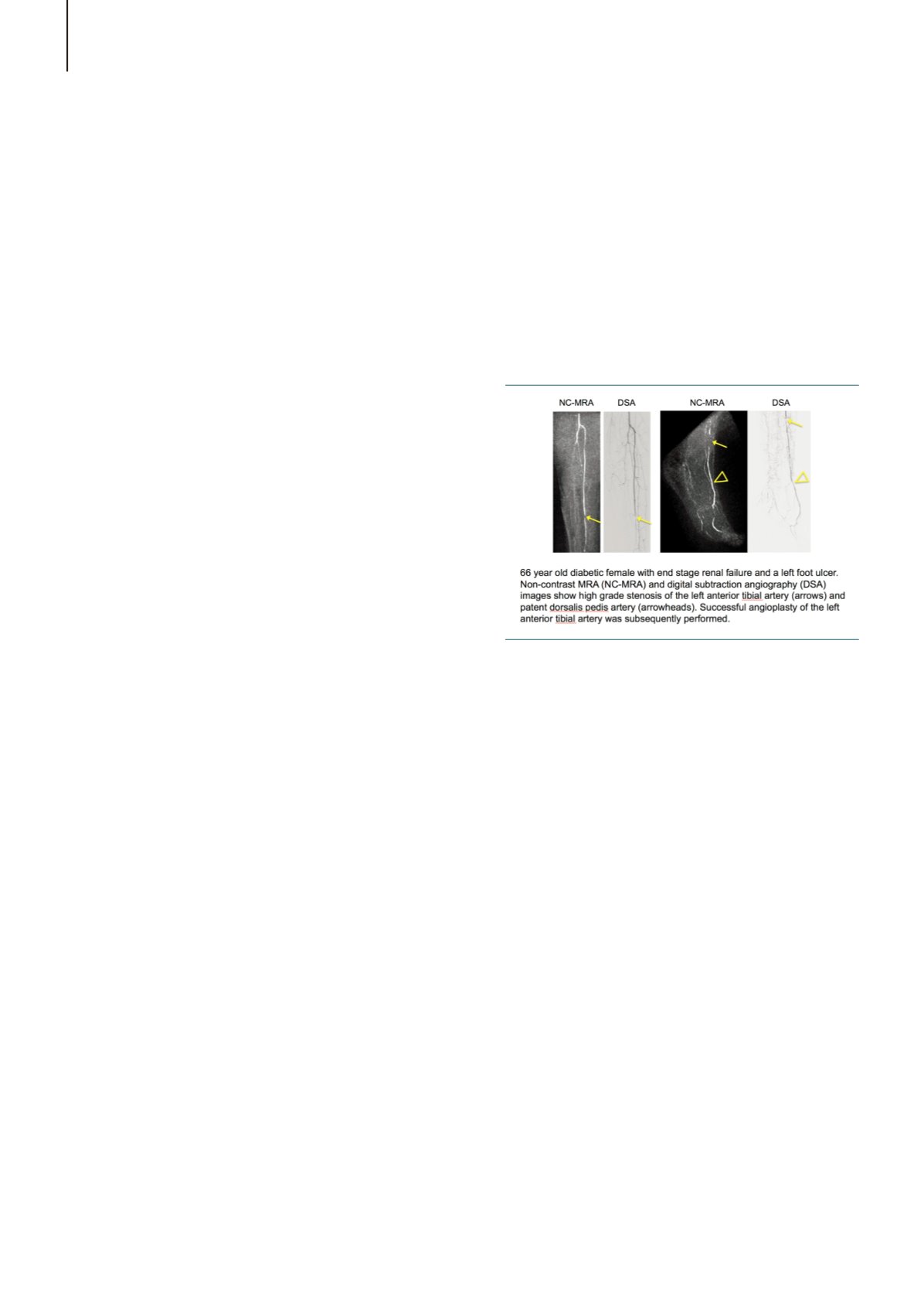
Our results were interesting. 28 patients
completed the study—one could
not fit completely within the MRI due
to morbid obesity, and one patient
could not complete the MRA due to
discomfort. Overall, the non-contrast
MRA was most accurate in assessing
the iliac and femoral vessels, with
accuracy decreasing for the more distal
vessels, particularly the pedal arteries.
However, readers felt more confident
in assessing the calf arteries with MRA
than with DSA, highlighting some of
the challenges of DSA, where the distal
vessels can be difficult to visualise due
to technical or patient factors. They were
least confident in interpreting imaging
of the feet, where vessels are small
and motion is common. As might be
expected, patients reported less overall
pain and anxiety with MRA compared
with DSA.
Results from different facets of the
project have been presented to our
peers at the Radiological Society of
North America, the International Society
for Magnetic Resonance in Medicine,
the Asian Society for Cardiovascular
Imaging, and shared with our vascular
surgery colleagues at the Australian
and New Zealand Society for Vascular
Imaging, with one manuscript submitted
and a second one in preparation.
Research is at times challenging and
results not something that are achieved
overnight. It is undoubtedly a team
effort, involving nursing, administrative,
radiography and medical staff. Not to
mention patients who kindly give their
time for the promise of improved care
for future patients. However, it is a
potentially educational and extremely
rewarding experience—apart from
myself, this project also provided
trainees with invaluable research
experience, of vital importance to the
future of our discipline.
This project would not have been
possible without the support of my
department and the College. I would
encourage anyone with an interesting
idea to take advantage of the
opportunity a research grant offers.
A/Prof Ruth Lim
Austin Health